Knee Muscle Anatomy Mri / Figure 12 From Normal Mr Imaging Anatomy Of The Knee Semantic Scholar. Weak adductor muscles may cause knee instability and adductor strain(2). These motions of the knee allow the body to perform such important movements as walking, running, kicking, and jumping. More images for knee muscle anatomy mri » Abnormal anatomy with normal signal, i.e. 9, popliteal a & v.
This tool is at the same time useful for the training and teaching of the anatomy, but also for experts to illustrate a course or an explanation of a pathology to a patient, in particular in the context of the ruptures of the cross ligaments or the lesions of the. Thigh muscles also protect neurovascular structures as they go through the proximal hip joint to the knee and lower leg(3). I designed musculoskeletal mri specifically with the radiology resident in mind but anyone is welcome to the site. Superiorly, it extends to the level of the crossing of the biceps femoris tendon, and remains superficial to fcl in this location.10 Anatomy basic knee mri checklist.
Diz Anatomisi Mri And Mrg Aksiyal from konez.com Magnetic resonance imaging is particularly well suited for the medical evaluation of the musculoskeletal (msk) system including the knee, shoulder, ankle, wrist and elbow. Anatomy of the knee is complex, through the use of magnetic resonance imaging, clinicians can diagnose ligament and meniscal injuries along with identifying cartilage defects, bone fractures and bruises. These motions of the knee allow the body to perform such important movements as walking, running, kicking, and jumping. Jul 03, 2018 · the muscles of the knee include the quadriceps, hamstrings, and the muscles of the calf. This tool is at the same time useful for the training and teaching of the anatomy, but also for experts to illustrate a course or an explanation of a pathology to a patient, in particular in the context of the ruptures of the cross ligaments or the lesions of the. Superiorly, it extends to the level of the crossing of the biceps femoris tendon, and remains superficial to fcl in this location.10 Oct 01, 2009 · mri traumatic changes. More images for knee muscle anatomy mri »
Jul 03, 2018 · the muscles of the knee include the quadriceps, hamstrings, and the muscles of the calf.
Injuries such as anterior cruciate ligament, meniscus and rotator cuff tears are all easily diagnosed when there is a firm understanding and knowledge of human anatomy. Use the mouse scroll wheel to move the images up and down alternatively use the tiny arrows (>>) on both side of the image to move the images. Magnetic resonance imaging is particularly well suited for the medical evaluation of the musculoskeletal (msk) system including the knee, shoulder, ankle, wrist and elbow. Anatomy basic knee mri checklist. These muscles work in groups to flex, extend and stabilize the knee joint. Abnormal anatomy with normal signal, i.e. These motions of the knee allow the body to perform such important movements as walking, running, kicking, and jumping. This tool is at the same time useful for the training and teaching of the anatomy, but also for experts to illustrate a course or an explanation of a pathology to a patient, in particular in the context of the ruptures of the cross ligaments or the lesions of the. Weak adductor muscles may cause knee instability and adductor strain(2). There is a flat area of tendon originating from the knee. I designed musculoskeletal mri specifically with the radiology resident in mind but anyone is welcome to the site. Superiorly, it extends to the level of the crossing of the biceps femoris tendon, and remains superficial to fcl in this location.10 Each anatomical structure was labeled interactively.
Weak adductor muscles may cause knee instability and adductor strain(2). The medial thigh muscles are responsible for the adduction (movement of a body part toward the body's midline) of the leg. Use the mouse scroll wheel to move the images up and down alternatively use the tiny arrows (>>) on both side of the image to move the images. 7, semitendinosus muscle and tendon. Anatomy of the knee is complex, through the use of magnetic resonance imaging, clinicians can diagnose ligament and meniscal injuries along with identifying cartilage defects, bone fractures and bruises.
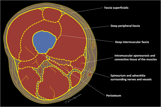
Fasciae Of The Musculoskeletal System Mri Findings In Trauma Infection And Neoplastic Diseases Insights Into Imaging Full Text from media.springernature.com These muscles work in groups to flex, extend and stabilize the knee joint. Anatomy arthrogram anatomy basic shoulder mri. Anatomy basic knee mri checklist. Injuries such as anterior cruciate ligament, meniscus and rotator cuff tears are all easily diagnosed when there is a firm understanding and knowledge of human anatomy. Weak adductor muscles may cause knee instability and adductor strain(2). 9, popliteal a & v. These motions of the knee allow the body to perform such important movements as walking, running, kicking, and jumping. Each anatomical structure was labeled interactively.
I designed musculoskeletal mri specifically with the radiology resident in mind but anyone is welcome to the site.
This tool is at the same time useful for the training and teaching of the anatomy, but also for experts to illustrate a course or an explanation of a pathology to a patient, in particular in the context of the ruptures of the cross ligaments or the lesions of the. The muscles of the knee include the quadriceps, hamstrings, and the muscles of the calf. I designed musculoskeletal mri specifically with the radiology resident in mind but anyone is welcome to the site. Thigh muscles also protect neurovascular structures as they go through the proximal hip joint to the knee and lower leg(3). 9, popliteal a & v. Each anatomical structure was labeled interactively. Superiorly, it extends to the level of the crossing of the biceps femoris tendon, and remains superficial to fcl in this location.10 Magnetic resonance imaging is particularly well suited for the medical evaluation of the musculoskeletal (msk) system including the knee, shoulder, ankle, wrist and elbow. More images for knee muscle anatomy mri » Abnormal anatomy with normal signal, i.e. Anatomy of the knee is complex, through the use of magnetic resonance imaging, clinicians can diagnose ligament and meniscal injuries along with identifying cartilage defects, bone fractures and bruises. Injuries such as anterior cruciate ligament, meniscus and rotator cuff tears are all easily diagnosed when there is a firm understanding and knowledge of human anatomy. The medial thigh muscles are responsible for the adduction (movement of a body part toward the body's midline) of the leg.
These motions of the knee allow the body to perform such important movements as walking, running, kicking, and jumping. 7, semitendinosus muscle and tendon. I designed musculoskeletal mri specifically with the radiology resident in mind but anyone is welcome to the site. Use the mouse scroll wheel to move the images up and down alternatively use the tiny arrows (>>) on both side of the image to move the images. Jul 03, 2018 · the muscles of the knee include the quadriceps, hamstrings, and the muscles of the calf.
2 from 9, popliteal a & v. 7, semitendinosus muscle and tendon. Anatomy arthrogram anatomy basic shoulder mri. Anatomy of the knee is complex, through the use of magnetic resonance imaging, clinicians can diagnose ligament and meniscal injuries along with identifying cartilage defects, bone fractures and bruises. These motions of the knee allow the body to perform such important movements as walking, running, kicking, and jumping. Anatomy basic knee mri checklist. Magnetic resonance imaging is particularly well suited for the medical evaluation of the musculoskeletal (msk) system including the knee, shoulder, ankle, wrist and elbow. Oct 01, 2009 · mri traumatic changes.
Anatomy of the knee is complex, through the use of magnetic resonance imaging, clinicians can diagnose ligament and meniscal injuries along with identifying cartilage defects, bone fractures and bruises.
Jul 03, 2018 · the muscles of the knee include the quadriceps, hamstrings, and the muscles of the calf. Anatomy arthrogram anatomy basic shoulder mri. Weak adductor muscles may cause knee instability and adductor strain(2). Use the mouse scroll wheel to move the images up and down alternatively use the tiny arrows (>>) on both side of the image to move the images. Magnetic resonance imaging is particularly well suited for the medical evaluation of the musculoskeletal (msk) system including the knee, shoulder, ankle, wrist and elbow. This mri knee sagittal cross sectional anatomy tool is absolutely free to use. 9, popliteal a & v. 7, semitendinosus muscle and tendon. I designed musculoskeletal mri specifically with the radiology resident in mind but anyone is welcome to the site. Injuries such as anterior cruciate ligament, meniscus and rotator cuff tears are all easily diagnosed when there is a firm understanding and knowledge of human anatomy. The medial thigh muscles are responsible for the adduction (movement of a body part toward the body's midline) of the leg. These muscles work in groups to flex, extend and stabilize the knee joint. Superiorly, it extends to the level of the crossing of the biceps femoris tendon, and remains superficial to fcl in this location.10
Share :
Post a Comment
for "Knee Muscle Anatomy Mri / Figure 12 From Normal Mr Imaging Anatomy Of The Knee Semantic Scholar"


Post a Comment for "Knee Muscle Anatomy Mri / Figure 12 From Normal Mr Imaging Anatomy Of The Knee Semantic Scholar"